Mr Ammar Alanbuki
Consultant Urological Surgeon
Specialist expertise: Andrology, Urology, Kidney Disease, Prostate, Urological Cancer, Erectile Dysfunction, Kidney Stones.
This process involves the taking of small cores of tissue from within the prostate to be examined by a pathologist for the presence or otherwise of prostate cancer

This process involves the taking of small cores of tissue from within the prostate to be examined by a pathologist for the presence or otherwise of prostate cancer
A prostate biopsy is usually suggested following an MRI scan of the prostate demonstrating risk areas within the gland, suspicious for prostate cancer. The MRI scan is usually performed if the PSA blood test is high, or if the prostate feels abnormal on examination.
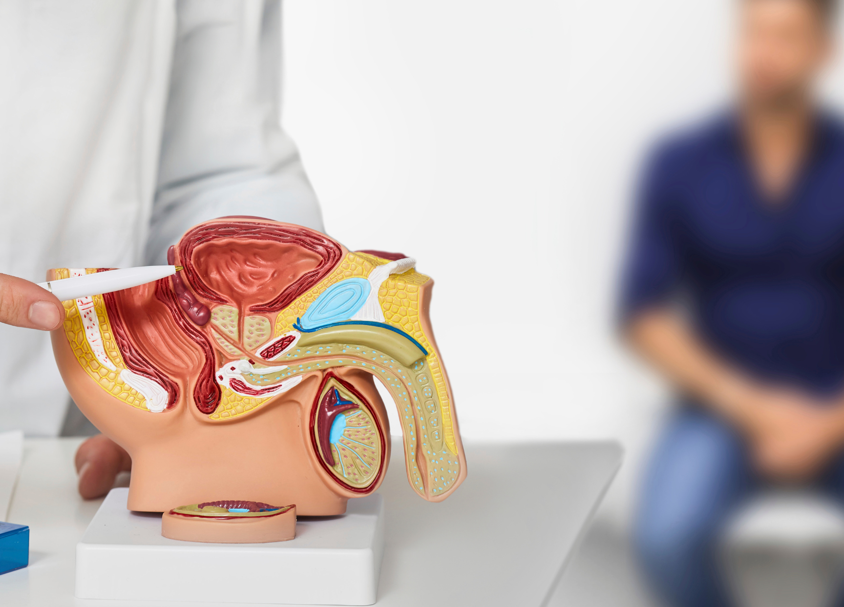
In the modern era, prostate biopsies are performed under local anaesthetic or sedation, using images of the prostate gained from an ultrasound probe placed up the back passage into the rectum. These images are used to guide the passage of a fine biopsy needle through the perineum (the area of skin between the scrotum and the anus) into the prostate to take the core of tissue. Prostate biopsies may be taken in a saturation fashion - sampling the whole prostate, or in a more targeted fashion based upon the MRI images. Fusion technology, that in real-time overlays the contour of the MRI scans onto the live ultrasound images, may be employed, particularly for smaller targets within the prostate, to improve accuracy.
Transperineal prostate biopsies are generally well tolerated with a rapid return to normal activity in a day or two. Associated risks include difficulty passing urine immediately after the procedure, perineal discomfort or pain and bruising, urinary infection and blood in the urine and semen.
Your urologist will have made arrangements to see you with the results of the biopsies. Should those biopsies prove positive for prostate cancer, the next steps are dictated by both the grade (how nice or nasty the cancer it is) and stage (how early or advanced it is) of the disease. Further investigations including PET scans, Bone scans or CT scans maybe required. If the biopsies are benign, but your PSA was raised, your urologist is likely to suggest a progarmme of PSA surveillance, looking to ensure stability over time.
Currently selected day
Available consultations
The specialists at OneWelbeck Men's Health use the latest innovations in healthcare to accurately diagnose and treat a wide range of urological conditions.