X-rays: How do they work and what are the risks?

X-rays provide the medical profession with a powerful diagnostic tool and can be utilised to help identify, diagnose, monitor, and even treat many pathologies.

What is an X-Ray?

An X-ray or radiograph has been a common imaging technique since Wilhelm Roentgen described his ‘invisible light’ in 1896. X-rays provide the medical profession with a powerful diagnostic tool and can be utilised to help identify, diagnose, monitor, and even treat many pathologies.

The exact method of using X-rays depends on the part of the body being examined. For example, a mammogram is specifically used to examine breast tissue, whereas a chest x-ray looks at the structures within your thorax.

Like many medical examinations, there are small risks associated with having an x-ray. However, in almost all cases the benefits greatly outweigh the risks. Your doctor will always assess the risks and benefits of each x-ray request.


What are X-Rays used for?

Although there are more sophisticated and more recently developed imaging techniques

(CT, MRI and Ultrasound to name a few), standard x-rays remain a fast and reliable way to identify many potential problems. These x-ray studies also use very tiny amounts of radiation compared to some other more advanced techniques like computed tomography (CT).

Some typical reasons your doctor may request an x-ray include:

  • To examine injuries to bones and joints
  • To monitor previously established abnormalities – typically monitoring the healing of a fracture
  • To assess effects of treatment
  • Breast screening
  • Heart size and shape
  • Lung problems
  • Tooth decay
  • Location of foreign bodies

What to expect and how to prepare

The examination is painless and fast. Indeed, you will be completely unaware that the x-ray has been completed. Typically, you are not required to do anything to prepare for an x-ray. Depending on that part of the body being imaged it may be helpful to wear loose comfortable clothing without fasteners or zips. Alternatively, you may be asked to change into a hospital gown and remove any metallic items such as jewellery, piercings and  watches before having an x-ray. It is important to make the imaging team operating the equipment aware of any metal implants from previous surgery.

Sometimes an x-ray study requires the use of a special ‘contrast’ agent beforehand. Contrast is used to provide clearer images of certain parts of your anatomy. Depending on what is being imaged contrast may be administered in a variety of ways:

  • As a liquid which is swallowed (such as a barium swallow)
  • As an injection into your blood vessels (such as a contrast enhanced CT)

Contrast typically contains either iodine or barium, both are dense elements that show up well on x-rays. It is important to let your doctor know if you have previously had a reaction to any medically administered contrast media – this is rare but recognised.

In some cases, for example, when imaging your stomach or intestines, your medical team may ask you to fast for a certain amount of time beforehand. During this period of fasting, you must avoid eating, you may also be asked to limit or avoid drinking certain liquids.

How are X-Rays taken?

X-rays are routinely performed by radiographers, radiologists, or other technicians in the radiology department of hospitals, specialised outpatient clinics or in dental practices.


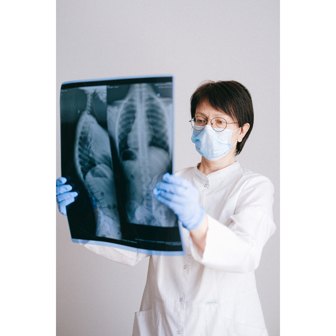
The medical professional operating the equipment will instruct you on how best to position yourself to produce a clear image. There are often a number of typical ‘standard’ views. For example an image of the chest is usually a single exposure, but an examination of an injured wrist may require two or even four different exposures. You may be asked to lie flat, sit or stand or adopt some other more specific positions to achieve these views depending on the body part being imaged.


A modern x-ray machine typically consists of an x-ray generating tube that emits the x-rays and a specialized plate containing x-ray sensors that capture the image. The appearance of x-ray machines can vary, some will be designed to work with you in the standing position, others are built above an examination bed to allow you to lie flat. Sometimes the equipment is be mounted on a large c-shaped arm to allow free movement around the body. However, all x-ray machines will position an x-ray tube on one side and the x-ray sensor plate on the other. This is because when the tube takes a ‘picture’ it emits x-rays which pass through the body. These x-rays will be absorbed differently depending on the nature of the tissue they have to pass through (bone, muscle, lung etc). This difference in absorption is what allows us to identify different tissues in the final image.


The most important thing to remember when having an x-ray taken is to remain as still as possible to allow clear crisp images to be captured. This may also involve briefly beathing in and holding your breath for chest or abdominal images. The whole process is usually very quick and will conclude once the team are happy with the image quality.


Risks and side effects

Modern x-ray machines now use a lot less radiation than in previous decades to produce high-quality images because of improvements in technology. Radiation occurs naturally and everyone on earth is exposed to a tiny amount of radiation every day. Natural radiation comes from several sources, radioactive material naturally occurs in tiny amounts in soil, water, rocks and even some plants. Cosmic radiation, which comes from outer space also partly consists of x-rays. This natural exposure is commonly referred to as background radiation. Although this naturally occurring radiation is not technically harmless, the levels are so low that any effects it has on the body are virtually undetectable. The Food and Drug Administration (FDA) in America found that a modern chest x-ray is equivalent to around 2.4 days of natural background radiation exposure.

Repeated exposure to large amounts of radiation (including x-rays) is associated with a small increase in your lifetime risk of cancer and so there is always a basic requirement to always use the minimum amount of radiation required to provide the information needed. This is the “As Low As Reasonably Achievable” (ALARA) principle. Indeed, the whole area of medical radiation use is governed by the IRMER (Ionising Radiation (Medical Exposure) Regulations) legislation. These are laws that have been put in place to maximise patient safety.


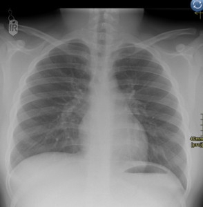
A normal chest x-ray

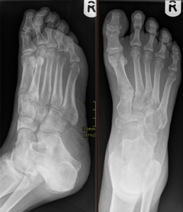
Two views of the right foot

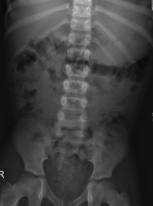
A normal abdominal x-ray showing the bones and the gas in the bowel.


How OneWelbeck
can help

Here at OneWelbeck, we have a team of top radiologists, state of the art facilities and diagnostics, and highly competitive financial packages for self-funding patients as well as those with private health care.

Imaging

Written by Professor Simon Padley

Professor Padley has a record in imaging research, extending over 30 years resulting in more than 130 peer reviewed publications based on imaging and interventional related subjects. He is a Professor of Practice in Diagnostic and Interventional Radiology in the Lung Division for the National Heart and Lung Institute, Imperial College, London.