Understanding the Jargon: Electrocardiograms (ECGs)

The ECG (electrocardiogram) is a widely used test that records the heart’s electrical activity. The ECG provides important information about the heart’s rhythm, and it can also help diagnose other heart problems.

What is an ECG?

The ECG (electrocardiogram) is a widely used test that records the heart’s electrical activity. The ECG provides important information about the heart’s rhythm, and it can also help diagnose other heart problems.

The heartbeat is regulated by electrical impulses that tell the heart muscle cells when to contract. The way in which these electrical impulses spread through the heart muscle determines the way in which the heart muscle contracts. This is known as the heart rhythm. As electrical impulses spread through the heart, the accompanying changes in electrical voltage can be measured by electrodes attached to the skin – the electrocardiogram.


How is an ECG performed?

The ECG is a simple test that usually takes 5-10 minutes to administer, and it does not involve the use of needles or x-rays. Ten stickers (‘electrodes’) are attached to the skin in specific places – one on each limb and six on the chest wall, overlying the heart. These electrodes are connected to an ECG machine by electrical cables, and a 10-second recording of the heart’s electrical activity is then made.


An example of a normal ECG

The ten electrodes attached to the skin are used by the ECG machine to create twelve ‘leads’, which view the heart’s electrical activity from different angles; this is reflected in the above example, which displays three separate lines at the top and a longer, single-lead ‘rhythm strip’ in the bottom line.


ECG terms: the different waves recorded by the ECG

As electrical impulses spread through the heart muscle, different ‘waves’ and ‘complexes’ are recorded by the ECG. This ECG shows a single beat of sinus rhythm – the normal heart rhythm. The first event is the ‘P wave’, which occurs as the electrical impulse spreads through the top chambers of the heart, the atria. The electrical pathway connecting the atria and the heart’s main pumping chambers (the ventricles) is very small, and it conducts electrical impulses relatively slowly, resulting in the flat line after the P wave. The electrical impulse then spreads very rapidly through the ventricles, resulting in the ‘QRS complex’. The electrical changes then return to their resting state as the heart relaxes and refills with blood for the next beat. This results in the ‘T wave’.


Other ECG terms: the PR Interval

The PR interval is measured from the start of the P wave to the onset of the QRS complex, and it reflects the time taken for electrical impulses to conduct from the atria to the ventricles. A short PR interval may be seen in a condition known as Wolff-Parkinson-White syndrome, where there is an extra electrical connection between the atria and ventricles. A long PR interval is described as first-degree atrioventricular block (also known as first-degree heart block).

The QRS complex is usually narrow, because the electrical impulse spreads rapidly through the ventricles via specialised tissue known as the conduction system: the His bundle, which then divides into the right and left bundle branches. If the electrical impulse cannot conduct down one or other of the bundle branches, the impulse takes longer to spread through the ventricle on the affected side, resulting in a prolonged QRS complex with a characteristic pattern described as either right or left bundle branch block. Fast heartbeats (tachycardias) may be described as either narrow-complex or broad-complex, depending on the width of the QRS complex during the tachycardia. Narrow-complex tachycardias arise from the atria, whereas broad-complex tachycardias may arise from either the atria or ventricles.


The QT Interval

The T wave occurs as the ventricles return to their resting state in preparation for the next heartbeat. Abnormalities of the T wave are seen in many heart conditions. The QT interval is measured from the start of the QRS complex to the end of the T wave. The QT interval is dependent upon the heart rate, and so a ‘correction’ that takes account of the heart rate is usually applied to the measurement in order to provide the Corrected QT interval (‘QTc’). A prolonged QT interval may be associated with a abnormal heart rhythms that arise from the ventricles.


Longer-term ECG monitoring

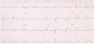
In certain situations, it is helpful to be able to monitor the ECG over an extended period. There are several ways to do this. An ambulatory ECG monitor (Holter monitor) is a portable monitor that attaches to the skin via sticky electrodes that record the ECG continuously, for periods ranging from 24 hours to two weeks. This is supplied with a symptom diary in which the date, time and nature of any symptoms occurring during the recording are entered. The symptom diary is extremely important, as it allows us to see exactly what is happening to the heart rhythm at the time that these symptoms occur. It is helpful to enter ‘no symptoms’ in the diary if none occurred whilst the monitor was being used, because many people have disturbed heart rhythms during ambulatory ECG monitoring, and many heart rhythm disturbances require treatment only if they are causing symptoms.

When symptoms that indicate a possible heart rhythm disturbance occur only infrequently, options for recording an ECG whilst these symptoms appear include the use of devices such as Kardia, which enables ECG recordings via Smartphones and tablets, or the Apple Watch. The Implantable Loop Recorder (ILR) is a small, battery-powered ECG that is implanted under the skin over the chest wall and records the heart rhythm continuously for up to three years. The ILR automatically stores ECG recordings of abnormal heart rhythms, and it also stores ECG recordings if the accompanying activator device is used when symptoms occur. The ILR can be monitored remotely by the hospital.


How OneWelbeck
can help

Here at OneWelbeck, we have a team of specialists, state of the art facilities and diagnostics, and highly competitive financial packages for self-funding patients as well as those with private health care.

Cardiology

Dr Simon Sporton

Written by Dr Simon Sporton, Consultant Cardiologist and Electrophysiologist at OneWelbeck Heart Health, specialising in the investigation and management of heart rhythm disorders.