
Dr Katie Lacy
Specialist expertise: Eczema, Psoriasis, Acne, Skin Rashes, Skin Cancer, Moles, Dermatology, General Dermatology, Adult Dermatology, Mole Checks, Mole Mapping, Cutaneous Immunology, Skin Cancer Surgery, Melanoma.
Dr Katie Lacy provides a detailed view on why mole mapping is important, plus a clinical examination for skin cancer surveillance.
The incidence and mortality from melanoma, the most serious type of skin cancer, continues to rise worldwide with around 16,700 cases per year in the UK. Since the 1990s the incidence rates have almost doubled and they are predicted to rise by around 10% between 2023- 2025. Melanoma is the 5th most common type of cancer in the UK, accounting for 4% of all new cancer cases [1]. Whilst there have been significant improvements in the management of advanced melanoma in the last few years, overall 25% of patients diagnosed with melanoma will go on to die from the disease. Early detection of melanoma at an early stage is therefore crucial as surgical removal of early stage disease usually results in cure. There is good evidence showing that adequate screening of individuals leads to higher pick-up rates of early-stage thin melanomas with a more favourable prognosis [2] and reduced medical costs [3,4].
Standard approaches to skin cancer diagnosis traditionally have relied on patients or Dermatologists to identify individual skin lesions of concern which may result in missed melanomas [5]. The use of mole mapping enables for early detection of melanoma by assessing for new and changing skin lesions - the most sensitive way of detecting melanomas at a stage where treatment confers excellent prognosis. The technique utilizes digital whole body imaging in addition to dermoscopy (a 10-15X magnified image of individual skin lesions providing detail on additional structures that cannot be seen by eye) to capture images of moles.
Sequential imaging at follow up visits allows for monitoring with great accuracy and identifies minor skin changes which may indicate early malignancy, allowing clinicians to detect subtle signs of in-situ (precancerous) melanoma or invasive melanoma with a higher sensitivity and specificity [6] before they are apparent to the naked eye/ to the patient. Sequential digital photography has also been shown to improve the detection rate of early-stage melanoma which may lack dermoscopic (magnified) features of melanoma [7-9] thus improving outcomes. In addition, it enables moles to be monitored over time for stability minimizing the need for unnecessary removal of multiple benign moles.
The use of photography for the management of patients at higher risk for melanoma with atypical (moles that are irregular in size, shape and colour) is further supported by the National Institute for Health and Care Excellence (NICE) guidelines for melanoma [10] that stipulate that clinically atypical melanocytic lesions that do not need excision at first presentation should have baseline photography and then subsequent review of the lesion in order to identify early signs of melanoma. Furthermore the NICE guidance also recommends the consideration of personalised follow-up for people who are at increased risk of melanomas (for example people with multiple moles including atypical moles, a previous melanoma, family history of melanoma). Mole mapping for individuals at risk of melanoma is therefore currently offered within Dermatology departments in the NHS.
2D total body photography has been used for many years to assist in skin cancer surveillance and is used both within the NHS and many private mole screening clinics. Taking 2D images involves taking multiple separate photographs of the patient standing in a variety of positions which can either overlap or fail to include moles if they are not captured on the specific anatomical angle required. It is a time consuming process often taking up to 30 minutes. Recent technological advances however have led to the development of 3D imaging which is designed to facilitate 360 degree rotation to view all body angles including curved surfaces that are often not captured adequately with 2D imaging. Early adopters of the technology worldwide have demonstrated the additive benefit of 3D image capture due to the capability of imaging almost the entire skin surface [11-12].
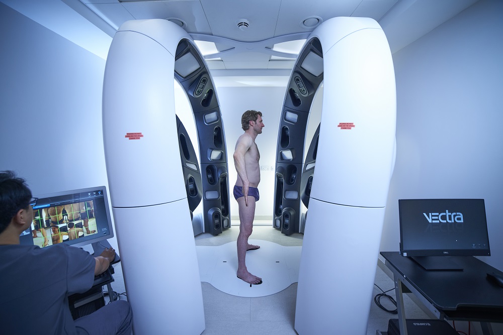
OneWelbeck are fortunate to have been able to acquire the state of the art VECTRA WB360 Whole Body 3 Dimensional Imaging system (Canfield Scientific)[13], the first of its kind in the UK. It uses 92 cameras to image the skin surface and stereophotogrammetry to construct a high-resolution 3D avatar of the patient. The patient stands in the device and image capture is taken within a matter of milliseconds- allowing for convenient, efficient and discrete acquisition of the required body images. In Australia, where the technology has been more widely adopted, a survey of over 1000 patients has indicated a high level of patient acceptance and confidence in the technology [14]. On subsequent visits with repeat imaging, usually recommended on an annual basis, an inbuilt AI system is able to identify new and changing skin lesions which is able to guide the clinician to areas on the skin requiring examination, reducing the risk of missing subtle early changes. The machine is able to detect changes in moles, new suspicious pigmented lesions in addition to development of non- melanoma skin cancers such as basal cell carcinomas. Patients are provided with a memory stick containing the photographic images obtained and are encouraged to use these to check for new and changing skin lesions in between appointments.
Ongoing research using the VECTRA 360 is currently underway to further develop the AI technology to allow for automated melanoma detection/ diagnosis, rather than relying on clinician examination of lesions. Researchers from the Memorial Sloan Kettering Cancer Centre in New York have recently demonstrated the possibility of automated melanoma detection using the machine [15] however larger studies are required to further evaluate this capability before its routine implementation.
In summary, 3D mole mapping allows for significantly enhanced monitoring of the skin in combination with physician and patient examination to allow for early detection and treatment of skin cancer.
Dr Katie Lacy is experienced in treating patients with general dermatological conditions including eczema, psoriasis, acne, skin rashes and cancerous and benign skin lesion. Dr Lacy also has extensive experience of the use of mole mapping and dermoscopy in order to identify suspect skin lesions that require removal.