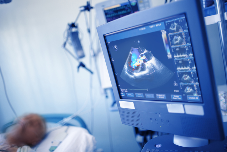
Heart Ultrasound Scans – What to Expect

Do you have a question about having a heart ultrasound? Here at OneWelbeck Heart Health we can provide answers and advice on what happens during a typical ultrasound scan.

What is a heart ultrasound (echocardiogram)?

Ultrasound scans of the heart, or echocardiograms, are essential imaging tests that help our experienced consultant cardiologists understand what’s going on inside your heart and the surrounding blood vessels.

They are used in the analysis of the function and structure of your heart and are critical in the diagnosis of certain heart conditions, including cardiomyopathy. 

You may need to have a heart ultrasound if you are experiencing symptoms that suggest you may have a problem with your heart, have already been diagnosed with a heart condition or to check your heart health before or after surgery. 

You might feel nervous about having a heart ultrasound scan for the first time if you don’t know what to expect. Let’s put your mind at ease by taking a look at what happens during the test, the different kinds of echocardiograms and why you may need one. 

Heart Ultrasound Scans - OneWelbeck


What to expect from a heart ultrasound scan

When we want to understand what is going on inside the heart, imaging tests are essential. One of the most common tests carried is a heart ultrasound; known more commonly as an echocardiogram. When someone has an echocardiogram carried out, it helps doctors analyse how the heart is working and that everything is performing as expected.

You might feel slightly nervous about having a heart ultrasound scan carried out for the first time if you don’t know what to expect. Let’s discuss the test, what happens during the examination, and what information a doctor expects to get from a heart ultrasound.


Different types of echocardiograms and what they involve

There are several ways an echocardiogram can be carried out, but a transthoracic echocardiogram (TTE) is the most common type. 

At OneWelbeck, echocardiograms are carried out by our expert sonographers, who report the findings of the scan to our consultant cardiologists.

Echocardiograms are performed in our outpatient clinic, so you will be able to go home or back to work shortly after. Most echocardiograms take between 15 and 60 minutes. 

The type of echocardiogram you will need depends on your symptoms or the heart condition being assessed. Your cardiologist will advise you which type is most appropriate. 

Heart Ultrasound Scans - OneWelbeck


Transthoracic echocardiogram (TTE)

To have a TTE, you'll be asked to remove your clothes from your waist up and put on a hospital gown before lying on a medical bed on your left side. 

Your sonographer will attach several sticky electrodes (sensors) to your chest. These are then attached to an electrocardiograph (ECG) monitor, which records your heart’s electrical activity during the test.

The sonographer will then apply a lubricating gel to your chest or directly to the ultrasound probe. They will glide the probe over your chest, and images of your heart will appear on a screen in real time. 

You might hear a swishing noise during the scan, which is the sound of your blood flowing through your heart.

A TTE is non-invasive and there is no preparation required for this test. 

Heart Ultrasound Scans - OneWelbeck


A transoesophageal echocardiogram (TOE)

A transoesophageal echocardiogram is less commonly used than a TTE but gives a more detailed view of your heart and valves as it collects pictures from inside your chest, rather than outside.

It is a minimally invasive procedure where your consultant carefully passes a thin, flexible ultrasound probe down your throat, into your oesophagus (food tube), which is right next to your heart. It can give a clearer view of your heart than a TTE as the ultrasound signal doesn’t have to travel through the bones and tissues in your chest.

Your throat will be numbed with a local anaesthetic spray and you will also be given a sedative to help you relax during the procedure. Your doctor may ask you to stop eating and drinking for a few hours before this test.

Heart Ultrasound Scans - OneWelbeck


A stress echocardiogram

A stress echocardiogram is the same as a TTE but is carried out during or just after a period of exercise on a treadmill or static bike. This gives your cardiologist an impression of how well your heart performs and functions when stressed and looks at the blood supply of the heart if you have chest pain. 

If you’re unable to exercise, you may be given a medication to make your heart work harder instead.

Your doctor may ask you to stop eating and drinking for a few hours before the test and to not smoke or drink caffeine for 24 hours before.

Heart Ultrasound Scans - OneWelbeck


A contrast echocardiogram

In some cases, your sonographer may inject a dye, called contrast, into your bloodstream before a TTE. This dye helps create a clear image of your heart on the ultrasound screen. 

A bubble echocardiogram

A bubble echocardiogram, also called a bubble saline contrast echocardiogram, uses an injection of saline mixed with your blood to form small bubbles to identify a hole in the heart called a patent foramen ovale (PFO).

The saline is injected into a vein in the back of your hand and as the bubbles move through your bloodstream and into your heart, they will be tracked using an echocardiogram.

There is no preparation required for this type of echocardiogram.

Heart Ultrasound Scans - OneWelbeck


What problems can a heart ultrasound scan identify?

An echocardiogram may be used for many reasons including:

  • A general health check of the heart

  • Identifying the cause of cardiac symptoms, such as chest pain or palpitations

  • Diagnosis of heart conditions, especially if you have a genetic risk factor


An echocardiogram can be used to detect and diagnose:

  • Congenital heart disease

  • Cardiomyopathy

  • Endocarditis

  • Pericardial disease

  • Valve disease and abnormalities

  • A cardiac tumour

  • An enlarged heart

  • Aortic aneurysm

  • Blood clots


How is a heart ultrasound done?

A probe is used to send sound waves from your chest to the heart and back. It is entirely harmless, and patients won’t feel a thing when the probe is moved around the chest.

It is common for the doctor to move the probe around slightly while checking the heart as it helps build a complete picture of how different parts of the heart work. The pictures received come as an “echo”, hence the term.


What does a patient do during a heart ultrasound?

In most instances, a patient will be asked to remove clothes from the upper body and then lie on their left side.


Would you be expected to exercise during an echocardiogram?

Sometimes doctors will want to understand how the heart works with stress. To do this they might ask the patient to run on the treadmill or to cycle on a static bike. You can find more information on that procedure.


How long does an ultrasound of the heart take?

A patient would expect their heart ultrasound appointment to last for 30 to 45 minutes, depending on how easy it is for doctors to get the images they need.


What does someone experience during a heart ultrasound?

The majority of patients won’t feel or notice anything while the ultrasound takes place. You will hear the sound of the flow of blood through the heart at times during the test when Doppler is used.


Do you need to take any specific medicine to have an echocardiogram carried out?

Usually, this test doesn’t need the patient to take any medicine. If the patient needs to have a bubble contrast echocardiogram, a saline injection will be needed.


Talk to echocardiogram specialists

If you have any questions about any of the tests carried out, or would like to speak with someone about visiting OneWelbeck for a test, please visit our contact page.


How OneWelbeck
can help

Here at OneWelbeck, we have a team of specialists, state of the art facilities and diagnostics, and highly competitive financial packages for self-funding patients as well as those with private health care.

Cardiology

What does a patient do during a heart ultrasound?

In most instances, you will be asked to remove your clothes from your waist up and then lie on your left side. Unless you are having a stress echocardiogram, you do not need to do anything while the test is done.

In some cases, your cardiologist will need to assess and understand how your heart works when taxed. To do this, they might ask you to run on the treadmill or cycle on a static bike before your sonographer performs the echocardiogram.

Most types of echocardiograms do not require you to take any medication. However, if you need a stress echocardiogram but cannot exercise, you may be given an injection to make your heart work harder. If you need a bubble contrast echocardiogram you will be given an injection of saline.

At OneWelbeck, your sonographer will report the findings of your echocardiogram to your cardiologist on the same day, who will talk you through the findings and any next steps.