Earliest availability 19 Feb
Dr Ozan Demir
Consultant Cardiologist
Specialist expertise: Interventional Cardiology, Hypertension, Cholesterol, Coronary Artery Disease, Preventative Cardiology, Heart Valve Disease, Cardiology.
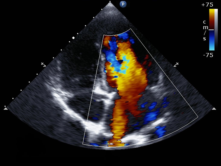
An echocardiogram (echo) is an ultrasound scan of the heart and nearby blood vessels to assess their structure and function.

02036532005
An echocardiogram is an ideal diagnostic test for anyone who wishes to have a general heart check. Additionally, if you have a family history of heart disease, if you have risk factors for cardiac disease, or cardiac symptoms such as chest pain or palpitations, we recommend you get an echocardiogram to ascertain the cause.
Your GP may also recommend having an echocardiogram.
For an echocardiogram, you’ll be offered a chaperone, asked to removed clothing covering the upper half and given a gown open at the front. You’ll be asked to lie on the couch. Three small stickers will be attached to your chest to monitor your heart rate and rhythm during the test. You usually will be asked to lie on the left side. This is to improve the image quality of the echocardiogram. Images will be taken by placing the ultrasound gel and probe on your chest.
An echocardiogram can detect:
There are two other variations of echocardiogram available at OneWelbeck Heart Health:
A stress echocardiogram carried out during exercise or after being given and injection of medication which makes the work harder. It is used to detect coronary artery disease and can show if there is narrowing of the blood vessels supplying the heart.
Bubble echocardiogram or bubble study
A bubble echocardiogram is carried out to detect holes between the top chambers of the heart. An echocardiogram is performed whilst a small amount of saline is injected into the bloodstream through a cannula in the arm. The saline and blood are mixed to create microbubbles which can be seen to enter the right size of the heart; if they pass to the left side a hole in the heart is confirmed.
Currently selected day
Available consultations