Second-Trimester Anatomy Scan and Uterine Artery Dopplers
A second-trimester scan is performed when you're between 20 and 22 weeks pregnant to assess fetal development and health.

02036532008
What is a second-trimester anatomy scan?
When you reach the point of this scan, you’re halfway through your pregnancy. The second-trimester anatomy scan is recommended between 20 and 22 weeks of pregnancy. This mid-pregnancy scan is offered to everybody in pregnancy as part of the Fetal Anomaly Screening Programme (FASP).
One of the main purposes of the anatomy scan is to perform a detailed assessment of the baby’s anatomy to detect the presence of any abnormalities, as well as assess the location of the placenta, and ensure the amniotic fluid around the baby is within the normal range. You can also find out the sex of your baby, should you wish to.
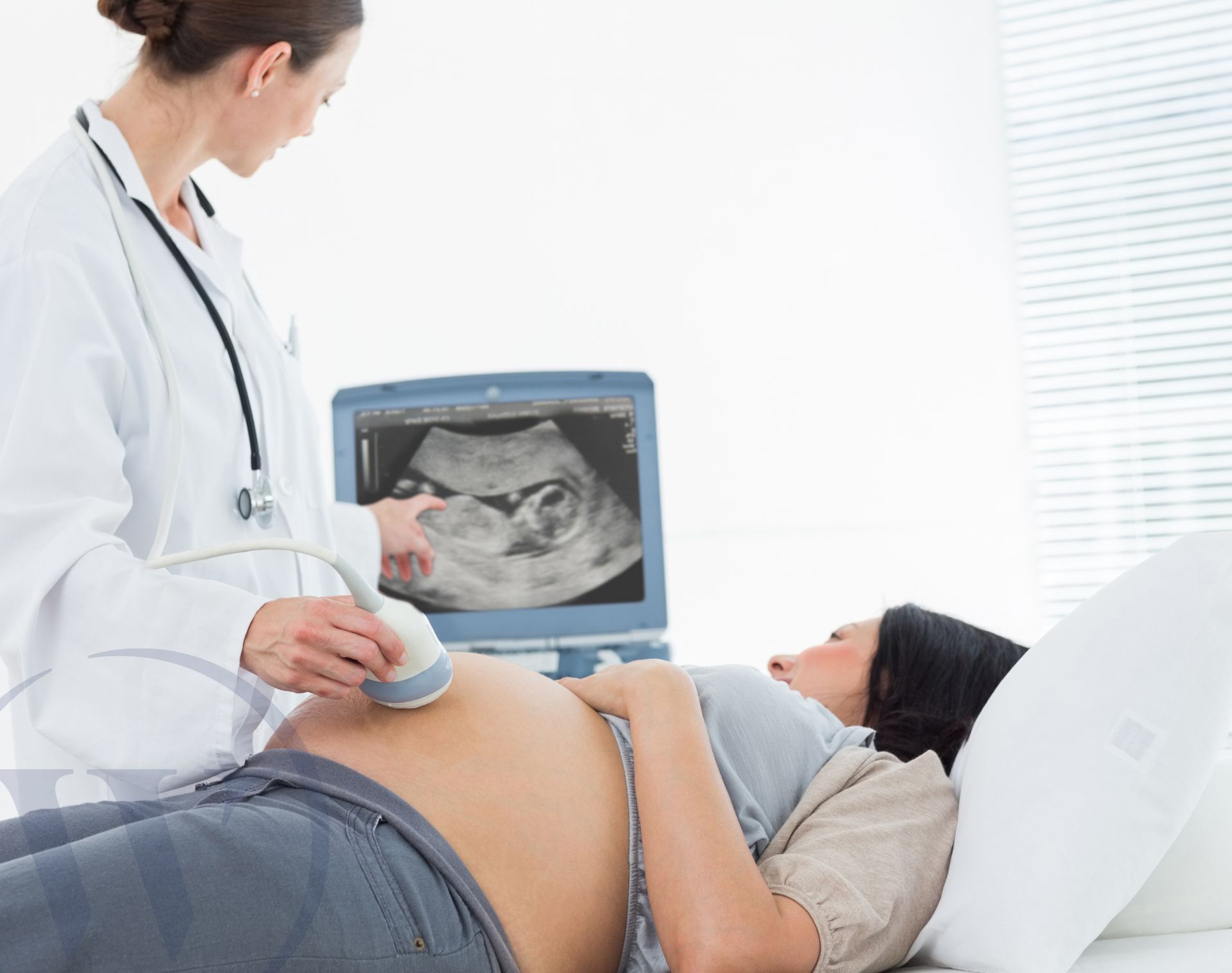
At Welbeck, second-trimester dating scans are performed in our state-of-the-art Women’s Health centre. The scan is done by one of our specialist sonographers, and you will be provided with a same-day report and results.
What is included in the anatomy check?
During the scan, we’ll perform a detailed assessment of the anatomy scan to assess your baby’s physical development and check for any potential structural defects or abnormalities. This involves taking measurements of your baby, including their head circumference, abdominal circumference, and femur length, which will be plotted in the detailed ultrasound report on a chart to show your baby’s overall growth.
The scan includes a thorough examination of the baby’s major organs, paying special attention to your baby’s:
- head and brain
- spine
- heart – assessing for any congenital heart defects
- face – any facial anomalies, for example cleft lip
- abdominal wall and internal organs – assessing the stomach, kidneys, and bladder (excluding abdominal wall defects)
- limbs – to ensure normal development of their arms and legs
Your sonographer will also perform uterine artery doppler studies at this scan – this simple and safe assessment is a quick examination to assess the blood flow in the arteries supplying the uterus (the womb), detecting any blood flow alterations. This provides further information on how the blood is flowing, if it’s flowing within the normal range (a low resistance reading), or if there is a high resistance reading – meaning the blood is having to work harder to get to the uterus.
A high resistance reading suggests there is an increased risk of having a smaller baby or developing a pregnancy condition known as pre-eclampsia. In the case of a high resistance reading, we will be able to guide you on the next steps and assist in appropriate referrals to formulate a management plan, which may include extra growth scans from 28 weeks.
In cases of a previous short cervix or any history of preterm delivery or concerns regarding premature delivery, a cervical assessment can be performed alongside this scan.
What is a cervical length assessment scan?
A cervical length assessment scan can be performed to estimate your risk of having a premature birth. It’s best to perform this from 16 weeks of pregnancy to measure the length of the cervix and identify any possible shortening.
Should you wish to add on this assessment, we will likely recommend performing it transvaginally (via your vagina), as this is usually the most accurate way to measure cervical length. This will be discussed with you at the time of the scan.
How is the scan performed?
This scan is performed over the tummy – a transabdominal scan. For a transabdominal ultrasound, ultrasound gel will be applied to your tummy so that an ultrasound probe can be moved back and forth. Live images will appear on a screen for you and your sonographer to see.
Should you wish to perform the cervical assessment, we may recommend doing this via transvaginal imaging if we feel it would be most accurate to measure the cervical length. This will be discussed with you at the time of the scan.
If you’re having a transvaginal (internal) ultrasound, you’ll be asked to lie on your back with your legs apart and knees bent. A small, lubricated probe will be gently inserted into your vagina and images will be produced on a screen. The probe will be fitted with a sterile, latex-free cover, and the probe will be moved slowly and gently to obtain images of your uterus and pelvis.
Is ultrasound safe in pregnancy?
There are no known risks to you or your baby from having an ultrasound scan, either with transabdominal or transvaginal imaging.
At Welbeck, we want to do everything we can to help you feel safe and comfortable in our care. If you would like, you can request a trained chaperone to attend the scan with you.
How do I prepare for the scan?
We recommend you attend your appointment with a full bladder. This allows optimal visualisation via transabdominal imaging. Should a different imaging technique and transvaginal scan be recommended, we will ask you to empty your bladder.
What happens after my scan?
After the scan, you’ll receive a comprehensive report discussing all the anatomy checked, along with the measurements performed and charted.
How much does a private second-trimester ultrasound scan cost?
At Welbeck, a second-trimester scan costs £250.
Why choose Welbeck?
It’s common to feel worried or anxious during pregnancy. At Welbeck, we offer expectant parents peace of mind through advanced fetal monitoring and are committed to supporting you at every stage of your pregnancy.
With access to colleagues across other specialties, we are also able to refer within the Welbeck ecosystem if needed to ensure you receive the best possible treatment as quickly as possible, all under one roof.
All appointments, testing, treatment, and follow-up appointments take place within our state-of-the-art facilities, enabling us to deliver accurate diagnostics and advanced treatments.
Booking a private early pregnancy ultrasound scan
If you’d like to book a private second-trimester ultrasound scan, simply get in touch with our Women’s Health centre to make an appointment.
FAQs
Will I receive Images?
Yes. Along with your report, we will provide you with printed images and hard copies for you to take home and share.
Can all anomalies be detected?
While ultrasound is an advanced and safe method of assessing the anatomy and providing the critical information we need is paramount – there’s unfortunately no test that can exclude all anomalies, and there are a few conditions and chromosomal or genetic conditions that may not be detected via ultrasound. Defects of the heart can be more challenging to detect and identify, with 50% of cardiac abnormalities being detectable. This is mind – most of the major cardiac abnormalities can be seen within this 50%. Our professional team are here to use our expertise, accuracy, and experience to exclude as many of the major anomalies as possible and ease your concerns.
What happens if an abnormality or concern is detected at the scan?
Should any abnormalities or concerns be detected on this scan, or if your sonographer requires a second opinion, we’ll recommend a repeat scan with one of our fetal medicine specialists here at Welbeck. We’ll discuss the scan findings with you at the time of the scan, and provide you with a copy of the full report for your records, which can also be shared with your midwife or obstetrician. Any recommendations for further testing referrals will be clearly and precisely communicated to you.
Do I still need the NHS scan?
Although we assess as much of the baby’s anatomy as possible at this point and provide reassurance in earlier gestation, we still recommend that you attend the NHS fetal anatomy scan as another safe way of checking the anatomy. This scan should be routinely offered by your NHS hospital, and we strongly recommend you have your scheduled scans if offered by your provider for your continuity of care.