Dr Anmol Malhotra
Director of the Breast Service & Consultant Radiologist
Specialist expertise: Breast Imaging, Photodynamic Therapy, Radiology, Breast Cancer, Reconstructive Breast Surgery.
If you are showing breast symptoms, your pathway will be different from regular breast screening.
Bookings: Tel: 

If you have found a lump or you are concerned about a new or unusual symptom in your breast or armpit, please contact us. We can arrange an appointment for you to see one of our breast surgeons, who can asses you and recommend the next steps which may include a mammogram and/or an ultrasound scan. Our administrative team can advise on costs if you do not have private health insurance but would like to self-pay.
Other forms of investigations and breast cancer detection include:
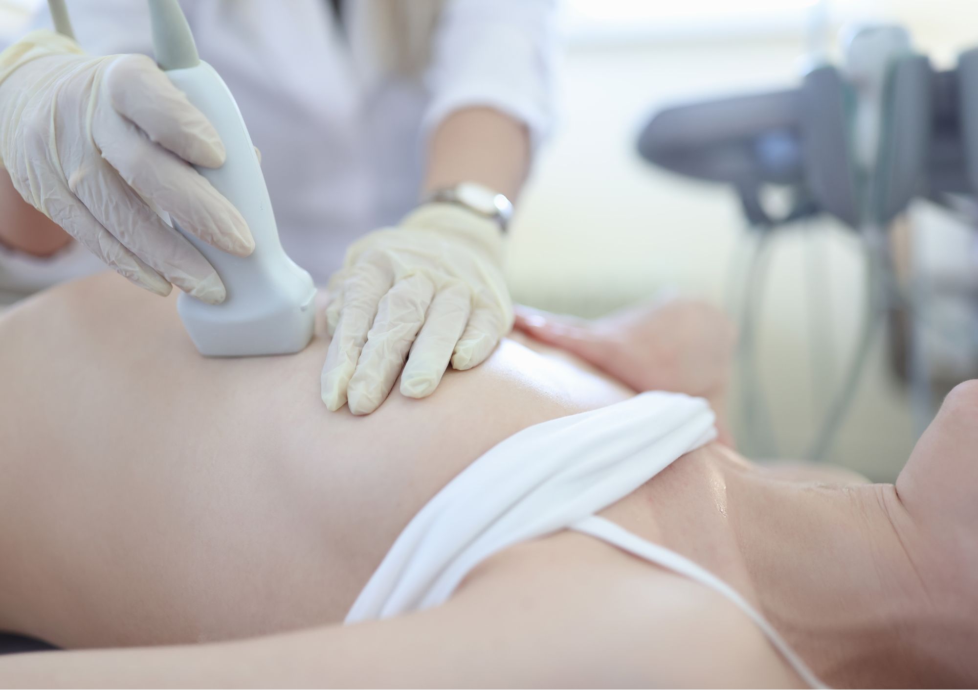
An ultrasound scan is performed when a change in the breast tissue is felt, or when an abnormality is detected on the mammogram. This modality is often performed in conjunction with mammography and can be used if the breast tissue is dense, or if the patient is under the age of 40.
The breast radiologist uses a probe that is slowly moved across the breast; high-frequency sound waves create a detailed image on a monitor. There are no known risks.
If the Consultant Radiologist feels it is necessary to take a tissue sample a needle test will be offered:
Your biopsy will be performed by the Radiologist usually using ultrasound or possibly mammography guidance. After local anaesthetic is injected using a very small needle into the overlying skin and then breast at the site of the sampling, the Radiologist uses the ultrasound images to guide a needle to the concerning area.
While a physical examination or imaging tests may suggest that breast cancer is present, tissue samples are ultimately needed to make the diagnosis, as well as determine the type of cancer and other characteristics.
Whichever type of imaging is utilized to guide the biopsy, the current standard of care at the completion of a biopsy is often the placement of a biopsy site marker. The marker is very small and the metal component is about the size of a sesame seed. This biopsy clip serves as a marker documenting where the tissue was sampled in the breast.
Ultrasound-guided breast biopsy does not involve exposure to ionizing radiation like x-ray (mammography) does. It is less invasive and usually leaves little or no scarring.
These samples are then sent to our Pathology lab for thorough and detailed examination. The results will be available usually within 48 hours, although this can be longer on occasion as sometimes the results have to be verified at a multidisciplinary team meeting.
This involves inserting a thin needle through the skin to collect a sample of cells or fluid. It is particularly helpful in distinguishing fluid-filled cysts from solid masses. This procedure is performed by the Radiologist using the ultrasound machine to guide them.
The vacuum-assisted biopsy-excision technique is performed under local anaesthesia using either ultrasound or mammography equipment.
It is a tissue sampling technique using imaging guidance to remove slightly larger samples of breast tissue through a single, small incision in the skin. The tissue sample is collected and sent to the laboratory for testing.
If you have recently been recalled via the NHS Breast Screening Program for further assessment but decide to have your assessment performed with private healthcare at Enhanced Breast Screening at OneWelbeck we can facilitate this either through self-pay or private healthcare insurance. With your permission, we will request your previous screening imaging via the Image Exchange Portal (IEP) prior to your visit. We can offer a full further assessment as clinically appropriate, using further tests such as tomosynthesis, magnification views, ultrasound scan, and needle biopsy if required. Biopsy results are usually available within 72 working hours.