The simple guide to MRIs and what they are used to diagnose

Magnetic resonance imaging (MRI) uses a large powerful magnet to produce a magnetic field. When combined with radio waves that are returned from the patient, the computer can produce amazingly detailed images of almost any part of the body. Like many imaging tests these pictures can help establish a diagnosis or assess changes over time.

Overview

Magnetic resonance imaging (MRI) uses a large powerful magnet to produce a magnetic field. When combined with radio waves that are returned from the patient, the computer can produce amazingly detailed images of almost any part of the body. Like many imaging tests these pictures can help establish a diagnosis or assess changes over time.

The MRI scanner is a large tube-shaped machine with a bed in the centre. The scanner is contained in a specially built room (or mobile unit). The patient lies on the bed, which can slide into the scanner to place the patient in the correct position and allow the study to be undertaken. The procedure usually takes about 20 minutes, and although noisy is otherwise straightforward, requiring no harmful radiation and is a completely painless and safe non-invasive examination.


How does the MRI scanner work

MRI scans are possible because the human body contains a large amount of water. Water molecules are made of hydrogen and oxygen atoms. In the core of the hydrogen atom is a small particle called a proton which acts like a small bar magnet. The powerful magnet of the MRI scanner can cause these protons to line up in one direction. The MRI scanner can then send small, targeted bursts of harmless radio waves to the part of the body being imaged. The radio waves bump the protons out of line. After a variable time (of less than a second) the protons flip back into line and at the same time will emit a burst of radio waves. It is these returning signals that an MRI scanner can detect. These tell the MRI computer the precise location of all the water molecules and also the sort of tissue that they are located in. Therefore bone, muscle, fat, fluid and damaged or abnormal tissue all have different signal characteristics, allowing a beautifully detailed image to be produced. What is more, these images can be produced in any orientation and can also be manipulated to accentuate certain characteristics of the tissues in question.

MRI allows your doctor to view your body in narrow slices, which can vary in thickness depending on the requirements of the study. In essence an MRI scan consists of multiple consecutive pictures of your body, just like slices in a loaf of bread. These slices can be made in any orientation depending on what your doctor is trying to see. Typically images are viewed from the patient’s feet, but are also produced from the sides (sagittal) or front (coronal) perspective.
Figure 1. Images of an MRI of the brain viewed from the feet (axial) side (sagittal) and front (coronal) point of view. Notice how the fluid in the eyes and around the brain is either grey or white depending on the MRI scan settings.

Figure 1. Images of an MRI of the brain viewed from the feet (axial) side (sagittal) and front (coronal) point of view. Notice how the fluid in the eyes and around the brain is either grey or white depending on the MRI scan settings.

Ultimately, these images allow the radiologist and the medical team to accurately detect even the smallest abnormalities and to differentiate between different types of normal and diseased tissue in areas of complex anatomy.


Why you might need an MRI

An MRI examination will be requested when detailed imaging is required to visual and assess internal organs, blood vessels, bones, joints and other tissues. When combined with the clinical examination and other special tests, a complete clinical assessment with allow the medical team to arrive at a likely diagnosis and guide subsequent investigations or treatments, to provide reassurance and monitor progress.


Common types of MRI examinations

MRI can be used to examine almost every area of the body and is particularly well suited to particular tasks. Since an MRI is quite a complex investigation, simpler tests will often be used initially, such as radiographs or ultrasound studies.

Below are a few examples of common types of MRI examination:
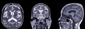
Brain and Spine

MRI scans are the most common method of imaging used for looking at the brain and spinal cord. Some of the principal conditions they can diagnoses in the brain and spinal cord are:

  • Aneurysms of the cerebral vessels
  • Strokes
  • Tumours
  • Multiple sclerosis
  • Spinal canal narrowing
  • Neurological disorders of brain and spine
  • Traumatic brain injury
  • Dementia

A specialist type of MRI called a functional MRI (fMRI) may be used to view the brain in action. An fMRI can be used to map brain activity and flow of blood in specific areas of the brain. This allows an accurate anatomical determination of which parts of the brain are carrying out essential functions, such as speech and movement. This can be an invaluable tool for some patients who may require brain surgery as it provides the surgeons with an understanding of an individual’s anatomy, including which side of the brain is undertaking important tasks such as speech and movement control.

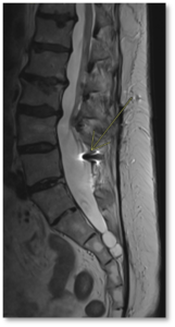
Figure 2. Image through the spine. The arrow is indicating an area of image disturbance caused by the previous surgery.

Heart and blood vessels

MRI scans of the heart and blood vessels can evaluate:

  • The thickness, movement, and health heart chambers
  • Pumping capacity and efficiency of the heart
  • Damage cause by previous heart attacks
  • Structural abnormalities of the heart or great vessels
  • Inflammation, blockages, or damage within blood vessels

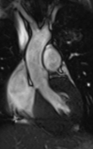
Figure 3. Coronal view of the left ventricle and the aorta – one frame from a cine acquisition series used to assess function.

Bones, joints, and muscles

MRI scans can be used to assess:

  • Problems with bones and joints, ligaments and muscles
  • Infections of the musculoskeletal system
  • Abnormalities caused by trauma or repetitive stress injuries

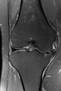
Figure 4. Image of the left knee viewed from the front showing damage to the medial part of the joint. This involves the cartilage disc in the joint, the articular cartilage lining of the joint and the bone.

Internal organs

MRI scans can also be used to assess for abnormalities, pathologies, and tumours in many other internal organs, such as:

  • Kidneys
  • Liver
  • Gallbladder and bile ducts
  • Spleen
  • Uterus
  • Ovaries
  • Pancreas
  • Bladder
  • Prostate

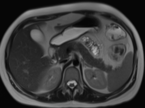
Figure 5. Image through the upper abdomen showing the liver, gall bladder, pancreas, spleen and kidneys.


How do I prepare for the scan?

You will be shown exactly where to lie on the scanner. You will be given some headphones to allow you to hear the technicians (and often listen to music or the radio to help pass the time). Once all preparations are complete the team will position the couch in the scanner. The bed may move throughout the scan and you will be given any instructions over the headphones and kept informed how much longer the study is likely to take and when to hold your breath (if needed).

The examination is painless and can last anywhere from 5 minutes to 60 minutes depending on the size of area being scanned. When the machine is scanning there may be a loud repetitive tapping or banging. This is completely normal – the headphones and/or disposable earplugs will reduce any noise to a comfortable level. Despite the noise the MRI contains no moving parts and there is no sensation related to the strong magnetic field.

Some types of study require contrast material. This is a fluid injected into a vein during the study to allow for better visualisation of some tissues.


What happens during the scan?

You will be shown exactly where to lie on the scanner. You will be given some headphones to allow you to hear the technicians (and often listen to music or the radio to help pass the time). Once all preparations are complete the team will position the couch in the scanner. The bed may move throughout the scan and you will be given any instructions over the headphones and kept informed how much longer the study is likely to take and when to hold your breath (if needed).

The examination is painless and can last anywhere from 5 minutes to 60 minutes depending on the size of area being scanned. When the machine is scanning there may be a loud repetitive tapping or banging. This is completely normal – the headphones and/or disposable earplugs will reduce any noise to a comfortable level. Despite the noise the MRI contains no moving parts and there is no sensation related to the strong magnetic field.

Some types of study require contrast material. This is a fluid injected into a vein during the study to allow for better visualisation of some tissues.


What happens after the test?

In most cases you will be able to resume your day as normal.


How do I get the results?

A specialist doctor called a radiologist will analyse your scan and produce a report which will be sent to your doctor. Your doctor will then contact you to discuss your results, either remotely or face to face. This whole process is quick and results should be with you in under a week.


Are there any risks?

MRIs are incredibly safe and extensive research has shown there are no associated health. Some patients may be sensitive to the contrast agent, and it is important to make you doctor aware pre-existing health conditions before the scan.
There are a few special circumstances that may exclude patients from having an MRI scan such as, cardiac pacemakers or prosthetic joints. It is important to inform your doctor if you are or may be pregnant.


How OneWelbeck
can help

Here at OneWelbeck, we have a team of top radiologists, state of the art facilities and diagnostics, and highly competitive financial packages for self-funding patients as well as those with private health care.

Imaging

Written by Professor Simon Padley

Professor Padley has a record in imaging research, extending over 30 years resulting in more than 130 peer reviewed publications based on imaging and interventional related subjects. He is a Professor of Practice in Diagnostic and Interventional Radiology in the Lung Division for the National Heart and Lung Institute, Imperial College, London.